Magnesium »
PDB 6ll8-6ly7 »
6lqk »
Magnesium in PDB 6lqk: Crystal Structure of Honeybee Ryr Ntd
Protein crystallography data
The structure of Crystal Structure of Honeybee Ryr Ntd, PDB code: 6lqk
was solved by
Y.Zhou,
L.Lin,
Z.Yuchi,
with X-Ray Crystallography technique. A brief refinement statistics is given in the table below:
| Resolution Low / High (Å) | 37.68 / 2.50 |
| Space group | P 21 21 21 |
| Cell size a, b, c (Å), α, β, γ (°) | 36.054, 75.363, 143.878, 90, 90, 90 |
| R / Rfree (%) | 22.3 / 28.8 |
Magnesium Binding Sites:
The binding sites of Magnesium atom in the Crystal Structure of Honeybee Ryr Ntd
(pdb code 6lqk). This binding sites where shown within
5.0 Angstroms radius around Magnesium atom.
In total only one binding site of Magnesium was determined in the Crystal Structure of Honeybee Ryr Ntd, PDB code: 6lqk:
In total only one binding site of Magnesium was determined in the Crystal Structure of Honeybee Ryr Ntd, PDB code: 6lqk:
Magnesium binding site 1 out of 1 in 6lqk
Go back to
Magnesium binding site 1 out
of 1 in the Crystal Structure of Honeybee Ryr Ntd
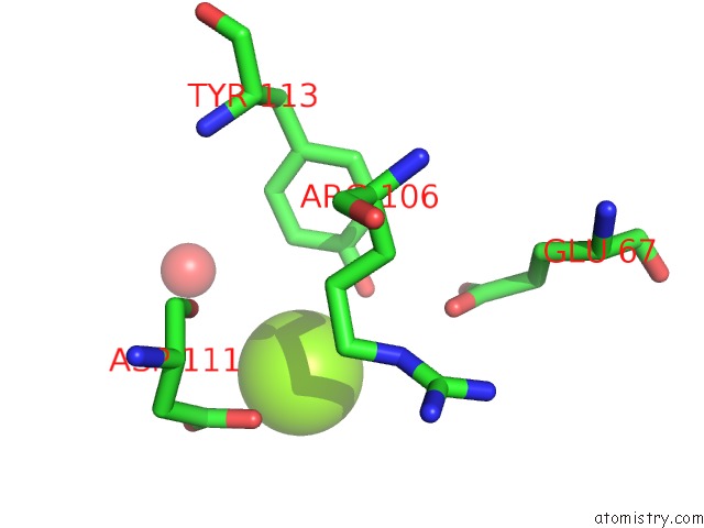
Mono view
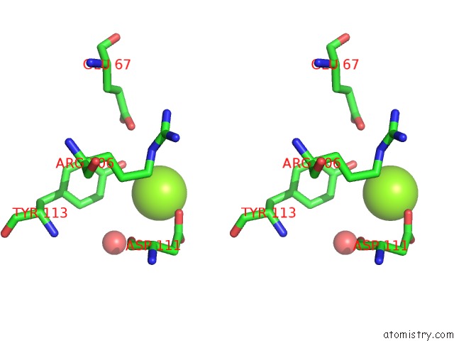
Stereo pair view
Mono view
Stereo pair view
A full contact list of Magnesium with other atoms in the Mg binding
site number 1 of Crystal Structure of Honeybee Ryr Ntd within 5.0Å range:
|
Reference:
Y.Zhou,
W.Wang,
N.M.Salauddin,
L.Lin,
M.You,
S.You,
Z.Yuchi.
Crystal Structure of the N-Terminal Domain of Ryanodine Receptor From the Honeybee, Apis Mellifera. Insect Biochem.Mol.Biol. V. 125 03454 2020.
ISSN: ISSN 0965-1748
PubMed: 32781205
DOI: 10.1016/J.IBMB.2020.103454
Page generated: Wed Aug 13 11:26:17 2025
ISSN: ISSN 0965-1748
PubMed: 32781205
DOI: 10.1016/J.IBMB.2020.103454
Last articles
Mg in 7DR0Mg in 7DR1
Mg in 7DU2
Mg in 7DSP
Mg in 7DSJ
Mg in 7DSI
Mg in 7DRP
Mg in 7DSH
Mg in 7DSA
Mg in 7DRX