Magnesium »
PDB 6ykx-6yxw »
6ysy »
Magnesium in PDB 6ysy: Skeletal Myosin Bound to Mph-220, Mgadp-VO4
Protein crystallography data
The structure of Skeletal Myosin Bound to Mph-220, Mgadp-VO4, PDB code: 6ysy
was solved by
L.Canon,
C.M.Kikuti,
M.Gyimesi,
A.Malnasi-Csizmadia,
A.Houdusse,
with X-Ray Crystallography technique. A brief refinement statistics is given in the table below:
| Resolution Low / High (Å) | 98.93 / 3.25 |
| Space group | P 21 21 21 |
| Cell size a, b, c (Å), α, β, γ (°) | 49.607, 119.424, 176.631, 90, 90, 90 |
| R / Rfree (%) | 21.8 / 23.2 |
Other elements in 6ysy:
The structure of Skeletal Myosin Bound to Mph-220, Mgadp-VO4 also contains other interesting chemical elements:
| Vanadium | (V) | 1 atom |
Magnesium Binding Sites:
The binding sites of Magnesium atom in the Skeletal Myosin Bound to Mph-220, Mgadp-VO4
(pdb code 6ysy). This binding sites where shown within
5.0 Angstroms radius around Magnesium atom.
In total only one binding site of Magnesium was determined in the Skeletal Myosin Bound to Mph-220, Mgadp-VO4, PDB code: 6ysy:
In total only one binding site of Magnesium was determined in the Skeletal Myosin Bound to Mph-220, Mgadp-VO4, PDB code: 6ysy:
Magnesium binding site 1 out of 1 in 6ysy
Go back to
Magnesium binding site 1 out
of 1 in the Skeletal Myosin Bound to Mph-220, Mgadp-VO4
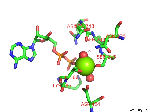
Mono view
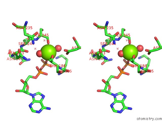
Stereo pair view
Mono view
Stereo pair view
A full contact list of Magnesium with other atoms in the Mg binding
site number 1 of Skeletal Myosin Bound to Mph-220, Mgadp-VO4 within 5.0Å range:
|
Reference:
M.Gyimesi,
A.I.Horvath,
D.Turos,
S.K.Suthar,
M.Penzes,
C.Kurdi,
L.Canon,
C.Kikuti,
K.M.Ruppel,
D.V.Trivedi,
J.A.Spudich,
I.Lorincz,
A.A.Rauscher,
M.Kovacs,
E.Pal,
S.Komoly,
A.Houdusse,
A.Malnasi-Csizmadia.
Single Residue Variation in Skeletal Muscle Myosin Enables Direct and Selective Drug Targeting For Spasticity and Muscle Stiffness. Cell V. 183 335 2020.
ISSN: ISSN 1097-4172
PubMed: 33035452
DOI: 10.1016/J.CELL.2020.08.050
Page generated: Wed Aug 13 21:19:50 2025
ISSN: ISSN 1097-4172
PubMed: 33035452
DOI: 10.1016/J.CELL.2020.08.050
Last articles
Mg in 7A17Mg in 7A0V
Mg in 6ZZX
Mg in 6ZXS
Mg in 7A0Q
Mg in 7A0P
Mg in 7A0C
Mg in 721P
Mg in 6ZXH
Mg in 6ZXG