Magnesium »
PDB 6ykx-6yxw »
6yt3 »
Magnesium in PDB 6yt3: Structure of the Mostonano Fusion Protein
Protein crystallography data
The structure of Structure of the Mostonano Fusion Protein, PDB code: 6yt3
was solved by
R.M.Benoit,
T.Bierig,
C.Collu,
S.Engilberge,
V.Olieric,
with X-Ray Crystallography technique. A brief refinement statistics is given in the table below:
| Resolution Low / High (Å) | 47.89 / 2.85 |
| Space group | H 3 2 |
| Cell size a, b, c (Å), α, β, γ (°) | 165.697, 165.697, 352.162, 90.00, 90.00, 120.00 |
| R / Rfree (%) | 23.3 / 26.3 |
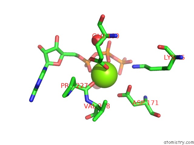
Magnesium Binding Sites:
The binding sites of Magnesium atom in the Structure of the Mostonano Fusion Protein
(pdb code 6yt3). This binding sites where shown within
5.0 Angstroms radius around Magnesium atom.
In total only one binding site of Magnesium was determined in the Structure of the Mostonano Fusion Protein, PDB code: 6yt3:
In total only one binding site of Magnesium was determined in the Structure of the Mostonano Fusion Protein, PDB code: 6yt3:
Magnesium binding site 1 out of 1 in 6yt3
Go back to
Magnesium binding site 1 out
of 1 in the Structure of the Mostonano Fusion Protein
Mono view
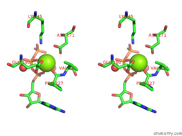
Stereo pair view
Mono view
Stereo pair view
A full contact list of Magnesium with other atoms in the Mg binding
site number 1 of Structure of the Mostonano Fusion Protein within 5.0Å range:
|
Reference:
G.Collu,
T.Bierig,
A.-S.Krebs,
S.Engilberge,
N.Varma,
R.Guixa-Gonzalez,
X.Deupi,
V.Olieric,
Poghosyan.E.,
R.M.Benoit.
Chimeric Single Alpha-Helical Domains As Rigid Fusion Protein Connections For Protein Nanotechnology and Structural Biology Biorxiv 2020.
DOI: 10.1101/2020.09.29.318410
Page generated: Wed Aug 13 21:20:06 2025
DOI: 10.1101/2020.09.29.318410
Last articles
Mg in 7A17Mg in 7A0V
Mg in 6ZZX
Mg in 6ZXS
Mg in 7A0Q
Mg in 7A0P
Mg in 7A0C
Mg in 721P
Mg in 6ZXH
Mg in 6ZXG