Magnesium »
PDB 8wp0-8x3y »
8wve »
Magnesium in PDB 8wve: Crystal Structure of Cyanobacterial Circadian Clock Protein Kaic
Protein crystallography data
The structure of Crystal Structure of Cyanobacterial Circadian Clock Protein Kaic, PDB code: 8wve
was solved by
Y.Furuike,
S.Akiyama,
with X-Ray Crystallography technique. A brief refinement statistics is given in the table below:
| Resolution Low / High (Å) | 47.14 / 2.73 |
| Space group | P 63 |
| Cell size a, b, c (Å), α, β, γ (°) | 94.188, 94.188, 179.168, 90, 90, 120 |
| R / Rfree (%) | 28 / 33.8 |
Magnesium Binding Sites:
The binding sites of Magnesium atom in the Crystal Structure of Cyanobacterial Circadian Clock Protein Kaic
(pdb code 8wve). This binding sites where shown within
5.0 Angstroms radius around Magnesium atom.
In total 4 binding sites of Magnesium where determined in the Crystal Structure of Cyanobacterial Circadian Clock Protein Kaic, PDB code: 8wve:
Jump to Magnesium binding site number: 1; 2; 3; 4;
In total 4 binding sites of Magnesium where determined in the Crystal Structure of Cyanobacterial Circadian Clock Protein Kaic, PDB code: 8wve:
Jump to Magnesium binding site number: 1; 2; 3; 4;
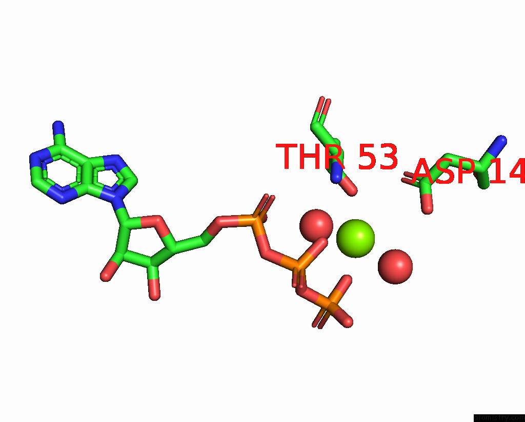
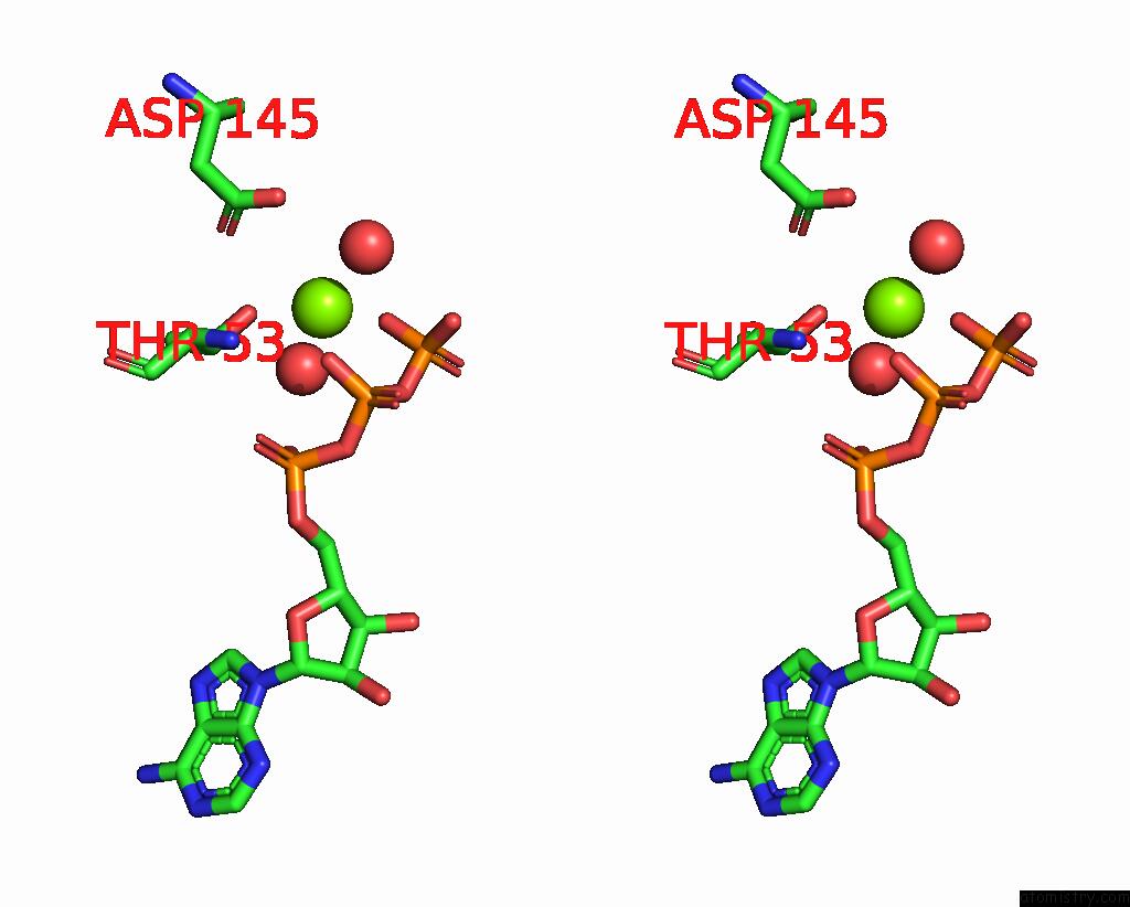
Magnesium binding site 1 out of 4 in 8wve
Go back to
Magnesium binding site 1 out
of 4 in the Crystal Structure of Cyanobacterial Circadian Clock Protein Kaic
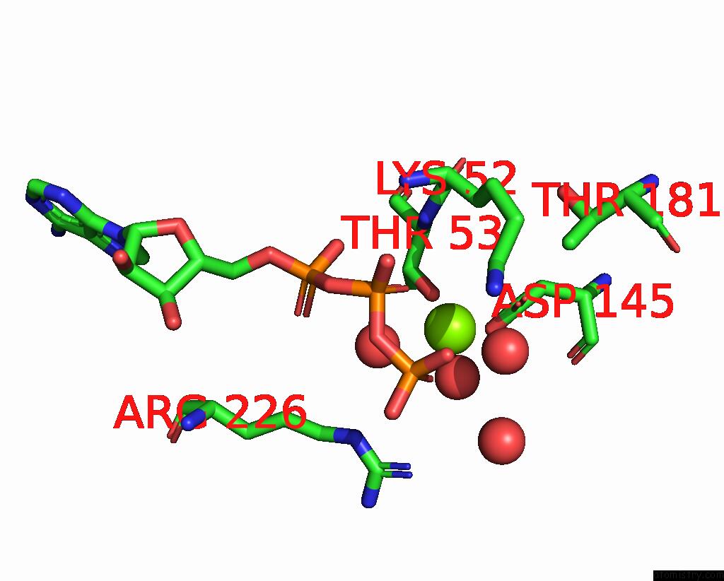
Mono view
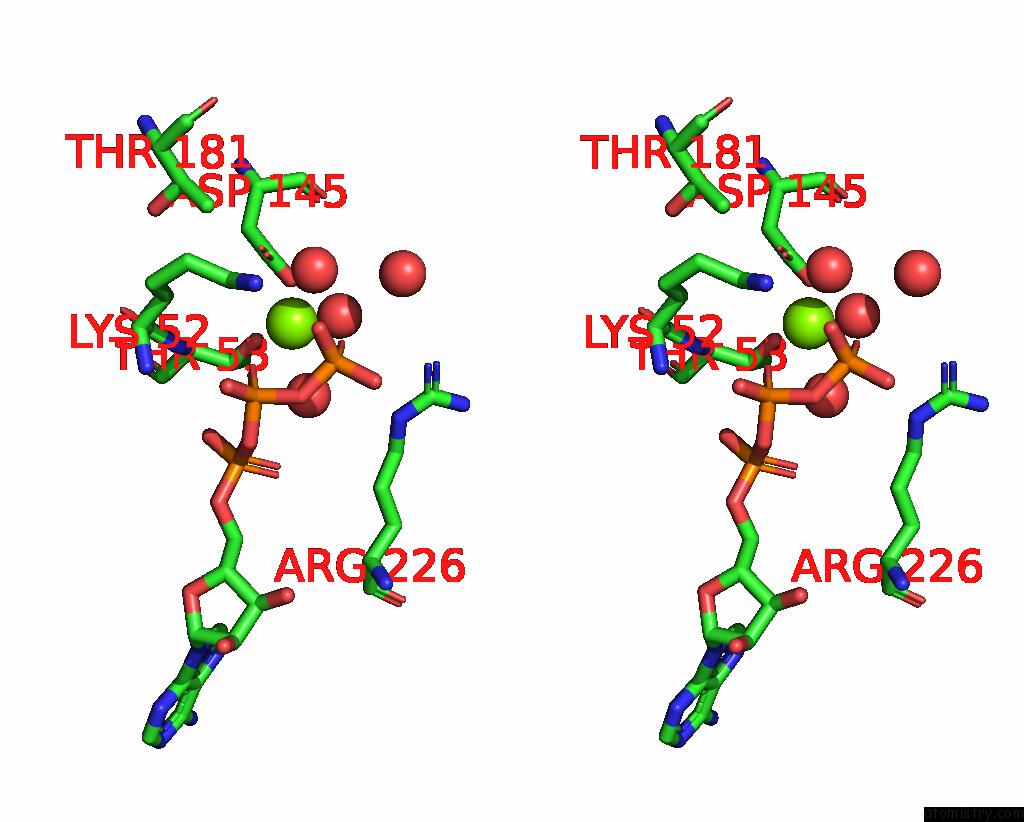
Stereo pair view
Mono view
Stereo pair view
A full contact list of Magnesium with other atoms in the Mg binding
site number 1 of Crystal Structure of Cyanobacterial Circadian Clock Protein Kaic within 5.0Å range:
|
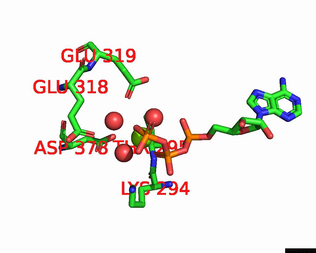
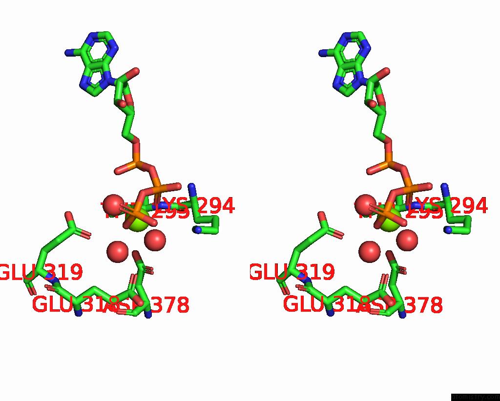
Magnesium binding site 2 out of 4 in 8wve
Go back to
Magnesium binding site 2 out
of 4 in the Crystal Structure of Cyanobacterial Circadian Clock Protein Kaic
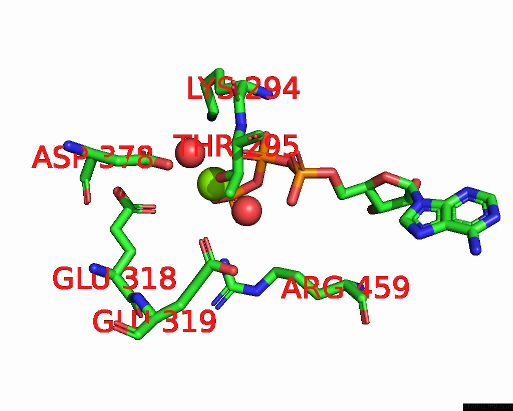
Mono view
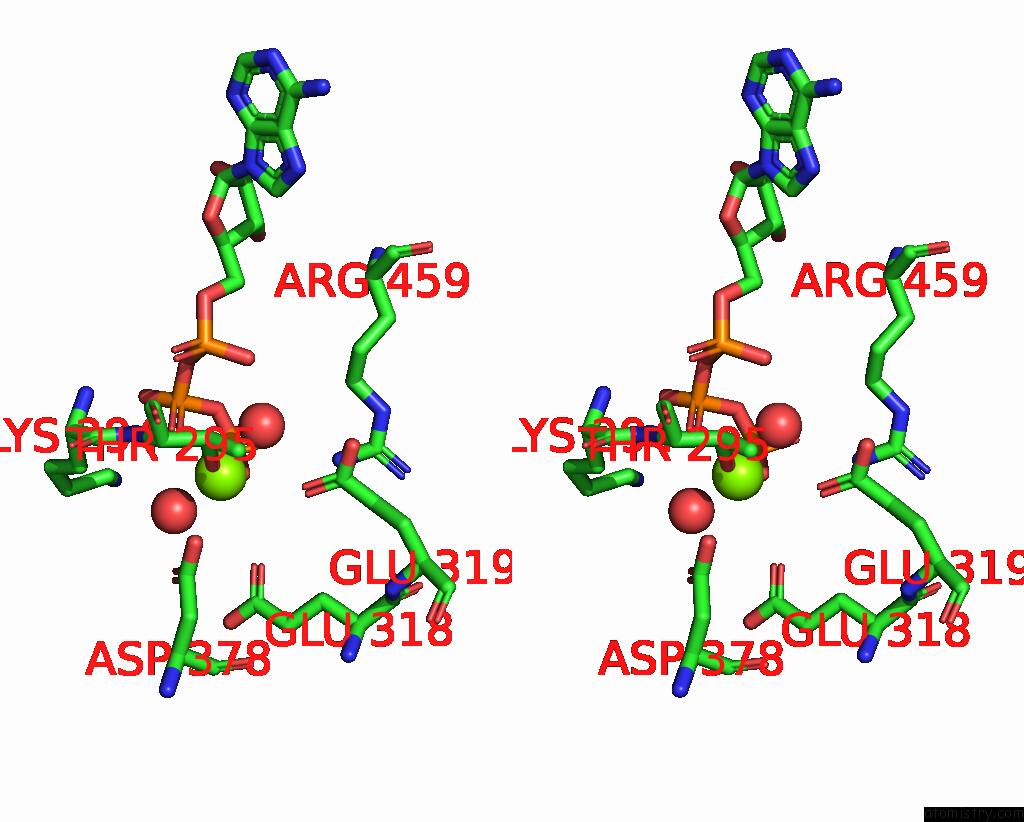
Stereo pair view
Mono view
Stereo pair view
A full contact list of Magnesium with other atoms in the Mg binding
site number 2 of Crystal Structure of Cyanobacterial Circadian Clock Protein Kaic within 5.0Å range:
|
Magnesium binding site 3 out of 4 in 8wve
Go back to
Magnesium binding site 3 out
of 4 in the Crystal Structure of Cyanobacterial Circadian Clock Protein Kaic
Mono view
Stereo pair view
Mono view
Stereo pair view
A full contact list of Magnesium with other atoms in the Mg binding
site number 3 of Crystal Structure of Cyanobacterial Circadian Clock Protein Kaic within 5.0Å range:
|
Magnesium binding site 4 out of 4 in 8wve
Go back to
Magnesium binding site 4 out
of 4 in the Crystal Structure of Cyanobacterial Circadian Clock Protein Kaic
Mono view
Stereo pair view
Mono view
Stereo pair view
A full contact list of Magnesium with other atoms in the Mg binding
site number 4 of Crystal Structure of Cyanobacterial Circadian Clock Protein Kaic within 5.0Å range:
|
Reference:
Y.Furuike,
E.Yamashita,
S.Akiyama.
Structure-Function Relationship of Kaic Around Dawn Biophys Physicobio. V. 20 2023.
ISSN: ESSN 2189-4779
DOI: 10.2142/BIOPHYSICO.BPPB-V21.0001
Page generated: Fri Aug 15 20:03:38 2025
ISSN: ESSN 2189-4779
DOI: 10.2142/BIOPHYSICO.BPPB-V21.0001
Last articles
Mg in 9G0DMg in 9G0C
Mg in 9G0A
Mg in 9G09
Mg in 9G08
Mg in 9FZF
Mg in 9FZ4
Mg in 9FYX
Mg in 9FYB
Mg in 9FXK