Magnesium »
PDB 1rtz-1s8f »
1s5g »
Magnesium in PDB 1s5g: Structure of Scallop Myosin S1 Reveals A Novel Nucleotide Conformation
Protein crystallography data
The structure of Structure of Scallop Myosin S1 Reveals A Novel Nucleotide Conformation, PDB code: 1s5g
was solved by
D.Risal,
S.Gourinath,
D.M.Himmel,
A.G.Szent-Gyorgyi,
C.Cohen,
with X-Ray Crystallography technique. A brief refinement statistics is given in the table below:
| Resolution Low / High (Å) | 43.44 / 3.10 |
| Space group | P 1 21 1 |
| Cell size a, b, c (Å), α, β, γ (°) | 84.051, 50.935, 161.590, 90.00, 98.36, 90.00 |
| R / Rfree (%) | 23.3 / 26.9 |
Other elements in 1s5g:
The structure of Structure of Scallop Myosin S1 Reveals A Novel Nucleotide Conformation also contains other interesting chemical elements:
| Calcium | (Ca) | 1 atom |
Magnesium Binding Sites:
The binding sites of Magnesium atom in the Structure of Scallop Myosin S1 Reveals A Novel Nucleotide Conformation
(pdb code 1s5g). This binding sites where shown within
5.0 Angstroms radius around Magnesium atom.
In total only one binding site of Magnesium was determined in the Structure of Scallop Myosin S1 Reveals A Novel Nucleotide Conformation, PDB code: 1s5g:
In total only one binding site of Magnesium was determined in the Structure of Scallop Myosin S1 Reveals A Novel Nucleotide Conformation, PDB code: 1s5g:
Magnesium binding site 1 out of 1 in 1s5g
Go back to
Magnesium binding site 1 out
of 1 in the Structure of Scallop Myosin S1 Reveals A Novel Nucleotide Conformation
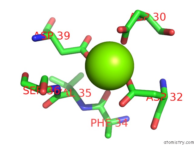
Mono view
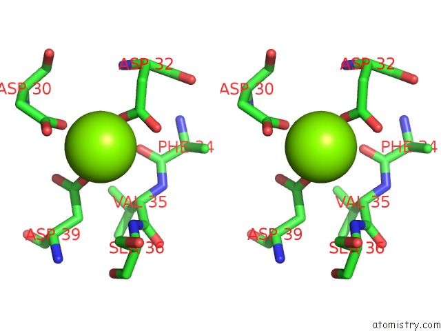
Stereo pair view
Mono view
Stereo pair view
A full contact list of Magnesium with other atoms in the Mg binding
site number 1 of Structure of Scallop Myosin S1 Reveals A Novel Nucleotide Conformation within 5.0Å range:
|
Reference:
D.Risal,
S.Gourinath,
D.M.Himmel,
A.G.Szent-Gyorgyi,
C.Cohen.
Myosin Subfragment 1 Structures Reveal A Partially Bound Nucleotide and A Complex Salt Bridge That Helps Couple Nucleotide and Actin Binding. Proc.Natl.Acad.Sci.Usa V. 101 8930 2004.
ISSN: ISSN 0027-8424
PubMed: 15184651
DOI: 10.1073/PNAS.0403002101
Page generated: Tue Aug 13 13:15:56 2024
ISSN: ISSN 0027-8424
PubMed: 15184651
DOI: 10.1073/PNAS.0403002101
Last articles
Mg in 1R0ZMg in 1R0X
Mg in 1R0A
Mg in 1R03
Mg in 1QZR
Mg in 1QU2
Mg in 1QYF
Mg in 1QVJ
Mg in 1QVI
Mg in 1QV9