Magnesium »
PDB 2jga-2mse »
2jk1 »
Magnesium in PDB 2jk1: Crystal Structure of the Wild-Type Hupr Receiver Domain
Protein crystallography data
The structure of Crystal Structure of the Wild-Type Hupr Receiver Domain, PDB code: 2jk1
was solved by
K.M.Davies,
E.D.Lowe,
C.Venien-Bryan,
L.N.Johnson,
with X-Ray Crystallography technique. A brief refinement statistics is given in the table below:
| Resolution Low / High (Å) | 40.23 / 2.10 |
| Space group | P 43 21 2 |
| Cell size a, b, c (Å), α, β, γ (°) | 89.931, 89.931, 53.874, 90.00, 90.00, 90.00 |
| R / Rfree (%) | 22.379 / 24.347 |
Magnesium Binding Sites:
The binding sites of Magnesium atom in the Crystal Structure of the Wild-Type Hupr Receiver Domain
(pdb code 2jk1). This binding sites where shown within
5.0 Angstroms radius around Magnesium atom.
In total only one binding site of Magnesium was determined in the Crystal Structure of the Wild-Type Hupr Receiver Domain, PDB code: 2jk1:
In total only one binding site of Magnesium was determined in the Crystal Structure of the Wild-Type Hupr Receiver Domain, PDB code: 2jk1:
Magnesium binding site 1 out of 1 in 2jk1
Go back to
Magnesium binding site 1 out
of 1 in the Crystal Structure of the Wild-Type Hupr Receiver Domain
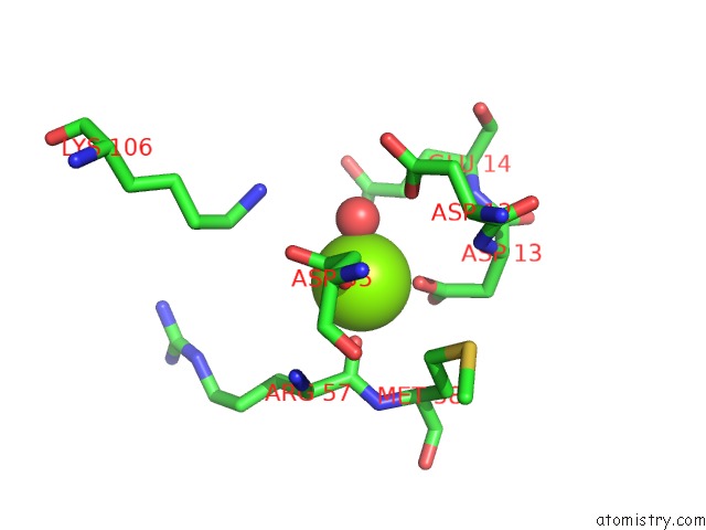
Mono view
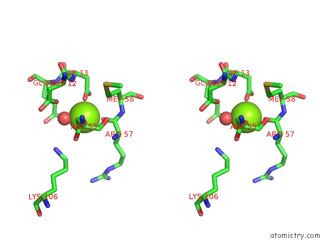
Stereo pair view
Mono view
Stereo pair view
A full contact list of Magnesium with other atoms in the Mg binding
site number 1 of Crystal Structure of the Wild-Type Hupr Receiver Domain within 5.0Å range:
|
Reference:
K.M.Davies,
E.D.Lowe,
C.Venien-Bryan,
L.N.Johnson.
The Hupr Receiver Domain Crystal Structure in Its Nonphospho and Inhibitory Phospho States. J.Mol.Biol. V. 385 51 2009.
ISSN: ISSN 0022-2836
PubMed: 18977359
DOI: 10.1016/J.JMB.2008.10.027
Page generated: Sun Aug 10 12:01:46 2025
ISSN: ISSN 0022-2836
PubMed: 18977359
DOI: 10.1016/J.JMB.2008.10.027
Last articles
Mg in 7A1AMg in 7A19
Mg in 6ZZY
Mg in 7A17
Mg in 7A0V
Mg in 6ZZX
Mg in 6ZXS
Mg in 7A0Q
Mg in 7A0P
Mg in 7A0C