Magnesium »
PDB 6m6b-6mmo »
6m8r »
Magnesium in PDB 6m8r: Crystal Structure of the KCTD16 Btb Domain in Complex with GABAB2 Peptide
Protein crystallography data
The structure of Crystal Structure of the KCTD16 Btb Domain in Complex with GABAB2 Peptide, PDB code: 6m8r
was solved by
S.Zheng,
A.C.Kruse,
with X-Ray Crystallography technique. A brief refinement statistics is given in the table below:
| Resolution Low / High (Å) | 38.64 / 3.20 |
| Space group | P 1 21 1 |
| Cell size a, b, c (Å), α, β, γ (°) | 91.964, 64.945, 114.137, 90.00, 99.79, 90.00 |
| R / Rfree (%) | 21.7 / 26.5 |
Magnesium Binding Sites:
The binding sites of Magnesium atom in the Crystal Structure of the KCTD16 Btb Domain in Complex with GABAB2 Peptide
(pdb code 6m8r). This binding sites where shown within
5.0 Angstroms radius around Magnesium atom.
In total 2 binding sites of Magnesium where determined in the Crystal Structure of the KCTD16 Btb Domain in Complex with GABAB2 Peptide, PDB code: 6m8r:
Jump to Magnesium binding site number: 1; 2;
In total 2 binding sites of Magnesium where determined in the Crystal Structure of the KCTD16 Btb Domain in Complex with GABAB2 Peptide, PDB code: 6m8r:
Jump to Magnesium binding site number: 1; 2;
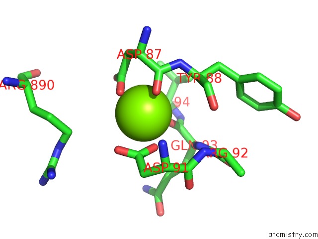
Magnesium binding site 1 out of 2 in 6m8r
Go back to
Magnesium binding site 1 out
of 2 in the Crystal Structure of the KCTD16 Btb Domain in Complex with GABAB2 Peptide
Mono view

Stereo pair view
Mono view
Stereo pair view
A full contact list of Magnesium with other atoms in the Mg binding
site number 1 of Crystal Structure of the KCTD16 Btb Domain in Complex with GABAB2 Peptide within 5.0Å range:
|
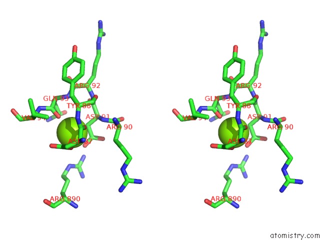
Magnesium binding site 2 out of 2 in 6m8r
Go back to
Magnesium binding site 2 out
of 2 in the Crystal Structure of the KCTD16 Btb Domain in Complex with GABAB2 Peptide

Mono view
Stereo pair view
Mono view
Stereo pair view
A full contact list of Magnesium with other atoms in the Mg binding
site number 2 of Crystal Structure of the KCTD16 Btb Domain in Complex with GABAB2 Peptide within 5.0Å range:
|
Reference:
S.Zheng,
N.Abreu,
J.Levitz,
A.C.Kruse.
Structural Basis For Kctd-Mediated Rapid Desensitization of Gababsignalling. Nature V. 567 127 2019.
ISSN: ISSN 0028-0836
PubMed: 30814734
DOI: 10.1038/S41586-019-0990-0
Page generated: Wed Aug 13 12:03:39 2025
ISSN: ISSN 0028-0836
PubMed: 30814734
DOI: 10.1038/S41586-019-0990-0
Last articles
Mg in 7DR0Mg in 7DR1
Mg in 7DU2
Mg in 7DSP
Mg in 7DSJ
Mg in 7DSI
Mg in 7DRP
Mg in 7DSH
Mg in 7DSA
Mg in 7DRX