Magnesium »
PDB 7pn9-7py0 »
7psx »
Magnesium in PDB 7psx: Structure of HOXB13 Bound to Hydroxymethylated Dna
Protein crystallography data
The structure of Structure of HOXB13 Bound to Hydroxymethylated Dna, PDB code: 7psx
was solved by
E.Morgunova,
A.Popov,
Y.Yin,
J.Taipale,
with X-Ray Crystallography technique. A brief refinement statistics is given in the table below:
| Resolution Low / High (Å) | 47.00 / 2.00 |
| Space group | P 1 |
| Cell size a, b, c (Å), α, β, γ (°) | 38.153, 55.541, 101.08, 88.02, 81.46, 84.94 |
| R / Rfree (%) | 26 / 28.3 |
Magnesium Binding Sites:
The binding sites of Magnesium atom in the Structure of HOXB13 Bound to Hydroxymethylated Dna
(pdb code 7psx). This binding sites where shown within
5.0 Angstroms radius around Magnesium atom.
In total 2 binding sites of Magnesium where determined in the Structure of HOXB13 Bound to Hydroxymethylated Dna, PDB code: 7psx:
Jump to Magnesium binding site number: 1; 2;
In total 2 binding sites of Magnesium where determined in the Structure of HOXB13 Bound to Hydroxymethylated Dna, PDB code: 7psx:
Jump to Magnesium binding site number: 1; 2;
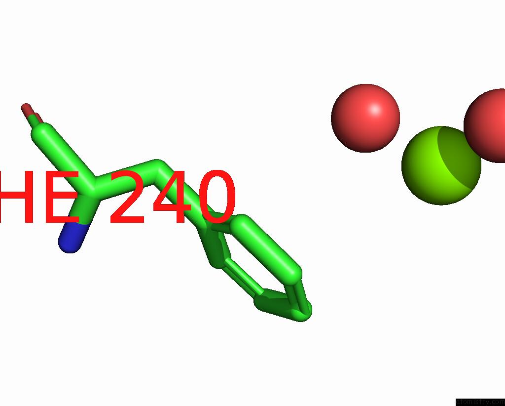
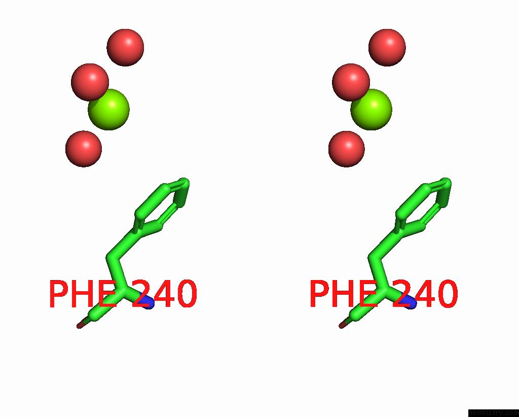
Magnesium binding site 1 out of 2 in 7psx
Go back to
Magnesium binding site 1 out
of 2 in the Structure of HOXB13 Bound to Hydroxymethylated Dna

Mono view

Stereo pair view
Mono view
Stereo pair view
A full contact list of Magnesium with other atoms in the Mg binding
site number 1 of Structure of HOXB13 Bound to Hydroxymethylated Dna within 5.0Å range:
|
Magnesium binding site 2 out of 2 in 7psx
Go back to
Magnesium binding site 2 out
of 2 in the Structure of HOXB13 Bound to Hydroxymethylated Dna
Mono view
Stereo pair view
Mono view
Stereo pair view
A full contact list of Magnesium with other atoms in the Mg binding
site number 2 of Structure of HOXB13 Bound to Hydroxymethylated Dna within 5.0Å range:
|
Reference:
E.Morgunova,
A.Popov,
Y.Yin,
J.Taipale.
Structure of HOXB13 Bound to Hydroxymethylated Dna To Be Published.
Page generated: Thu Oct 3 04:45:52 2024
Last articles
Mg in 5BJOMg in 5B8F
Mg in 5B6A
Mg in 5B4L
Mg in 5B4K
Mg in 5B3S
Mg in 5B48
Mg in 5B47
Mg in 5B46
Mg in 5B30