Magnesium »
PDB 8yo0-8z2l »
8z1r »
Magnesium in PDB 8z1r: Isocitrate Lyase MOMCL1
Protein crystallography data
The structure of Isocitrate Lyase MOMCL1, PDB code: 8z1r
was solved by
X.H.Liu,
W.H.Zhao,
with X-Ray Crystallography technique. A brief refinement statistics is given in the table below:
| Resolution Low / High (Å) | 32.81 / 1.82 |
| Space group | C 2 2 21 |
| Cell size a, b, c (Å), α, β, γ (°) | 118.43, 150.495, 128.243, 90, 90, 90 |
| R / Rfree (%) | 18.4 / 21.5 |
Magnesium Binding Sites:
The binding sites of Magnesium atom in the Isocitrate Lyase MOMCL1
(pdb code 8z1r). This binding sites where shown within
5.0 Angstroms radius around Magnesium atom.
In total 2 binding sites of Magnesium where determined in the Isocitrate Lyase MOMCL1, PDB code: 8z1r:
Jump to Magnesium binding site number: 1; 2;
In total 2 binding sites of Magnesium where determined in the Isocitrate Lyase MOMCL1, PDB code: 8z1r:
Jump to Magnesium binding site number: 1; 2;
Magnesium binding site 1 out of 2 in 8z1r
Go back to
Magnesium binding site 1 out
of 2 in the Isocitrate Lyase MOMCL1
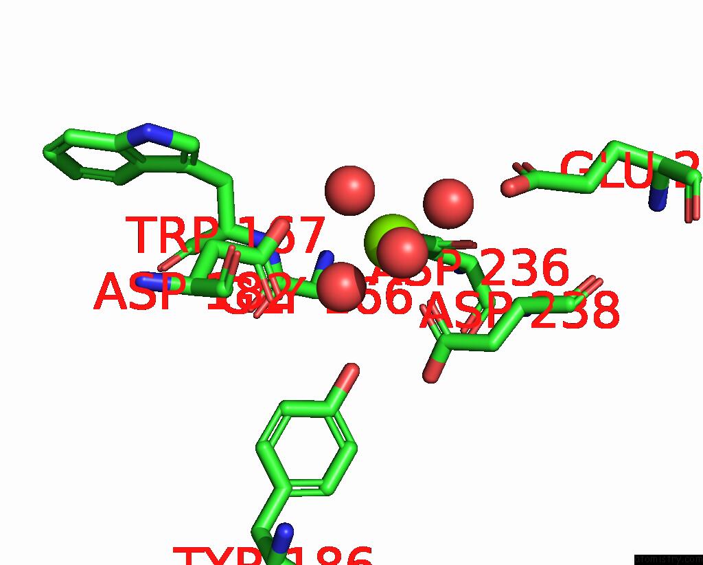
Mono view
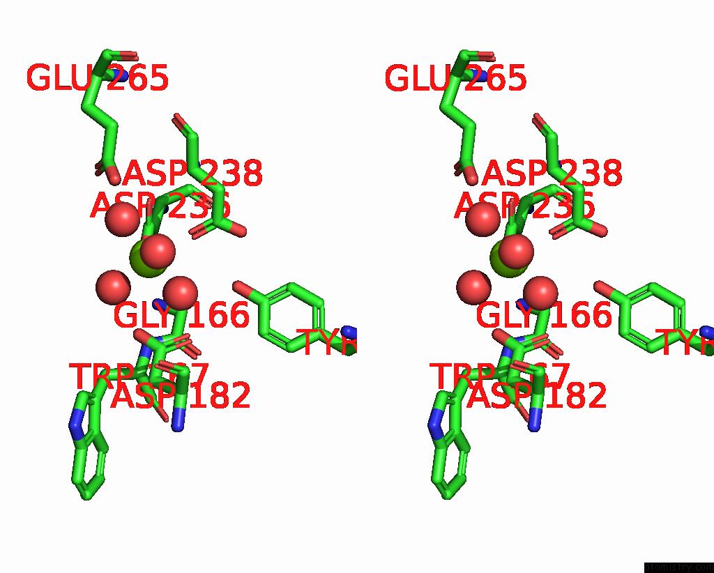
Stereo pair view
Mono view
Stereo pair view
A full contact list of Magnesium with other atoms in the Mg binding
site number 1 of Isocitrate Lyase MOMCL1 within 5.0Å range:
|
Magnesium binding site 2 out of 2 in 8z1r
Go back to
Magnesium binding site 2 out
of 2 in the Isocitrate Lyase MOMCL1
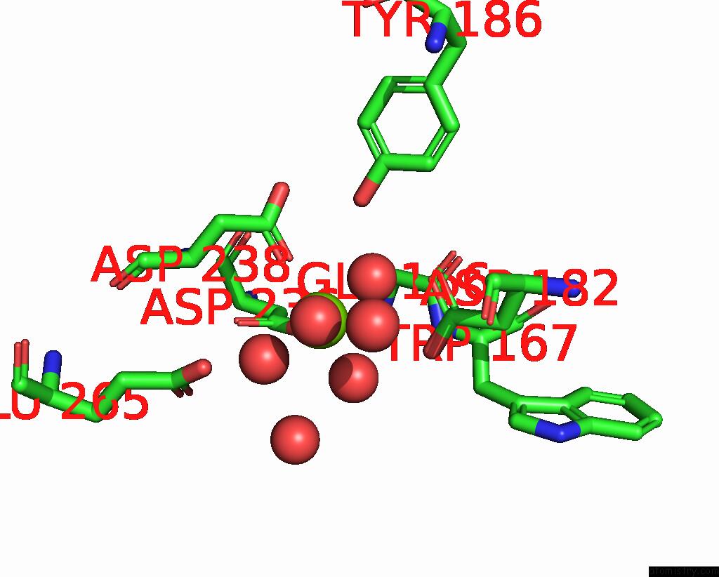
Mono view
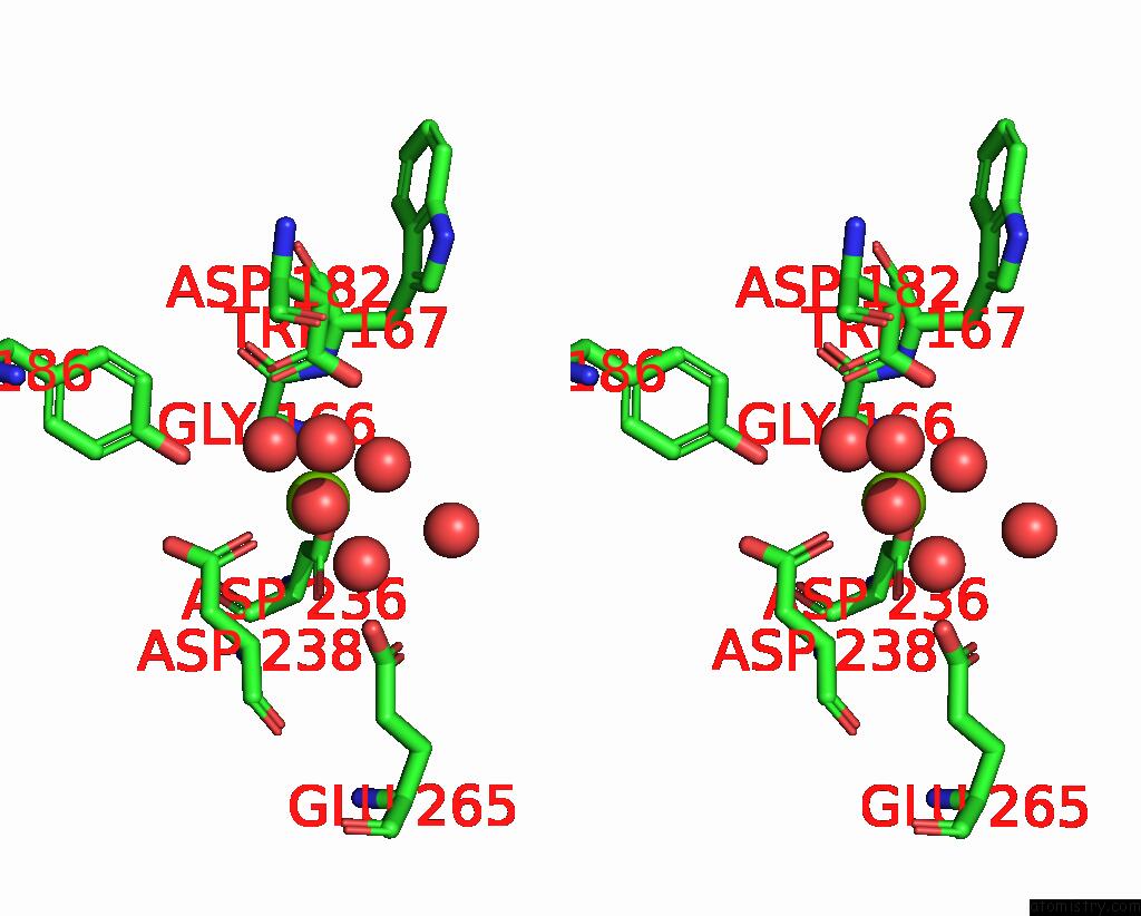
Stereo pair view
Mono view
Stereo pair view
A full contact list of Magnesium with other atoms in the Mg binding
site number 2 of Isocitrate Lyase MOMCL1 within 5.0Å range:
|
Reference:
X.H.Liu,
W.H.Zhao.
High-Resolution Crystal Structure of the MOMCL1 Protein To Be Published.
Page generated: Fri Aug 15 21:42:53 2025
Last articles
Mg in 9K3QMg in 9KOK
Mg in 9KBJ
Mg in 9KLN
Mg in 9KLQ
Mg in 9KK6
Mg in 9KI0
Mg in 9KHQ
Mg in 9KBI
Mg in 9KEV