Magnesium »
PDB 2gqr-2haw »
2h5o »
Magnesium in PDB 2h5o: Crystal Structure of Morange
Protein crystallography data
The structure of Crystal Structure of Morange, PDB code: 2h5o
was solved by
X.Shu,
S.J.Remington,
with X-Ray Crystallography technique. A brief refinement statistics is given in the table below:
| Resolution Low / High (Å) | 10.00 / 1.08 |
| Space group | P 21 21 21 |
| Cell size a, b, c (Å), α, β, γ (°) | 59.297, 62.353, 107.783, 90.00, 90.00, 90.00 |
| R / Rfree (%) | 14.4 / 19.1 |
Magnesium Binding Sites:
The binding sites of Magnesium atom in the Crystal Structure of Morange
(pdb code 2h5o). This binding sites where shown within
5.0 Angstroms radius around Magnesium atom.
In total only one binding site of Magnesium was determined in the Crystal Structure of Morange, PDB code: 2h5o:
In total only one binding site of Magnesium was determined in the Crystal Structure of Morange, PDB code: 2h5o:
Magnesium binding site 1 out of 1 in 2h5o
Go back to
Magnesium binding site 1 out
of 1 in the Crystal Structure of Morange
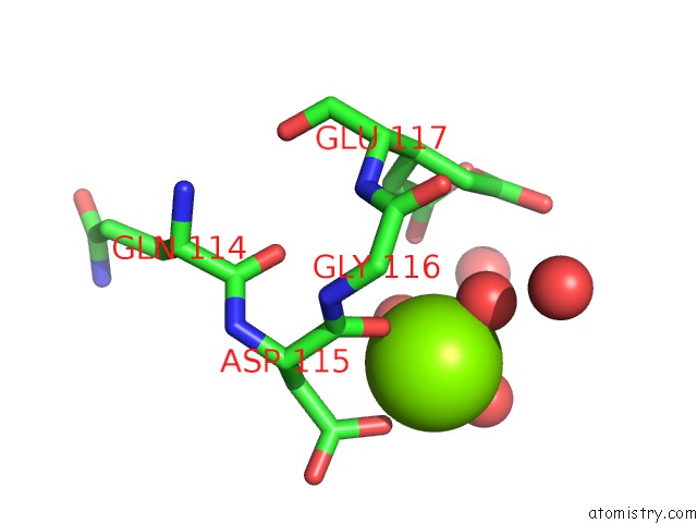
Mono view
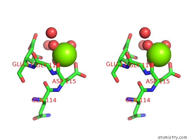
Stereo pair view
Mono view
Stereo pair view
A full contact list of Magnesium with other atoms in the Mg binding
site number 1 of Crystal Structure of Morange within 5.0Å range:
|
Reference:
X.Shu,
N.C.Shaner,
C.A.Yarbrough,
R.Y.Tsien,
S.J.Remington.
Novel Chromophores and Buried Charges Control Color in Mfruits Biochemistry V. 45 9639 2006.
ISSN: ISSN 0006-2960
PubMed: 16893165
DOI: 10.1021/BI060773L
Page generated: Sun Aug 10 11:17:46 2025
ISSN: ISSN 0006-2960
PubMed: 16893165
DOI: 10.1021/BI060773L
Last articles
Mg in 2R6OMg in 2R3B
Mg in 2R5T
Mg in 2R42
Mg in 2R20
Mg in 2R25
Mg in 2R2H
Mg in 2R23
Mg in 2R1X
Mg in 2R1W