Magnesium »
PDB 7kyc-7lc6 »
7l6s »
Magnesium in PDB 7l6s: Crystal Structure of the Atpase and Transducer Domains of Dna Topoisomerase II From Balamuthia Mandrillaris Cdc:V039: Baboon/San Diego/1986
Enzymatic activity of Crystal Structure of the Atpase and Transducer Domains of Dna Topoisomerase II From Balamuthia Mandrillaris Cdc:V039: Baboon/San Diego/1986
All present enzymatic activity of Crystal Structure of the Atpase and Transducer Domains of Dna Topoisomerase II From Balamuthia Mandrillaris Cdc:V039: Baboon/San Diego/1986:
5.6.2.2;
5.6.2.2;
Protein crystallography data
The structure of Crystal Structure of the Atpase and Transducer Domains of Dna Topoisomerase II From Balamuthia Mandrillaris Cdc:V039: Baboon/San Diego/1986, PDB code: 7l6s
was solved by
Seattle Structural Genomics Center For Infectious Disease (Ssgcid),
with X-Ray Crystallography technique. A brief refinement statistics is given in the table below:
| Resolution Low / High (Å) | 39.74 / 1.95 |
| Space group | C 2 2 21 |
| Cell size a, b, c (Å), α, β, γ (°) | 87.77, 187.25, 61.69, 90, 90, 90 |
| R / Rfree (%) | 16.7 / 19.6 |
Magnesium Binding Sites:
The binding sites of Magnesium atom in the Crystal Structure of the Atpase and Transducer Domains of Dna Topoisomerase II From Balamuthia Mandrillaris Cdc:V039: Baboon/San Diego/1986
(pdb code 7l6s). This binding sites where shown within
5.0 Angstroms radius around Magnesium atom.
In total only one binding site of Magnesium was determined in the Crystal Structure of the Atpase and Transducer Domains of Dna Topoisomerase II From Balamuthia Mandrillaris Cdc:V039: Baboon/San Diego/1986, PDB code: 7l6s:
In total only one binding site of Magnesium was determined in the Crystal Structure of the Atpase and Transducer Domains of Dna Topoisomerase II From Balamuthia Mandrillaris Cdc:V039: Baboon/San Diego/1986, PDB code: 7l6s:
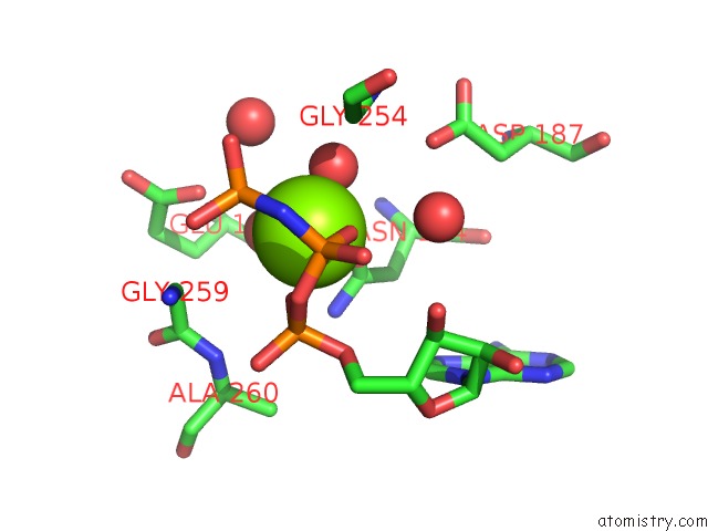
Magnesium binding site 1 out of 1 in 7l6s
Go back to
Magnesium binding site 1 out
of 1 in the Crystal Structure of the Atpase and Transducer Domains of Dna Topoisomerase II From Balamuthia Mandrillaris Cdc:V039: Baboon/San Diego/1986
Mono view

Stereo pair view
Mono view
Stereo pair view
A full contact list of Magnesium with other atoms in the Mg binding
site number 1 of Crystal Structure of the Atpase and Transducer Domains of Dna Topoisomerase II From Balamuthia Mandrillaris Cdc:V039: Baboon/San Diego/1986 within 5.0Å range:
|
Reference:
J.Abendroth,
D.Fox Iii,
D.D.Lorimer,
P.S.Horanyi,
T.E.Edwards.
Crystal Structure of the Atpase and Transducer Domains of Dna Topoisomerase II From Balamuthia Mandrillaris Lepto Id: Cdc:V039: Baboon/San Diego/1986 To Be Published.
Page generated: Wed Oct 2 23:18:21 2024
Last articles
Mg in 5TH3Mg in 5TIJ
Mg in 5THK
Mg in 5THX
Mg in 5TGK
Mg in 5TFG
Mg in 5TFF
Mg in 5TFJ
Mg in 5TFI
Mg in 5TDS